
How Do Humans Walk Stably on Two Legs? Two Developmental Shifts in the Embryonic Pelvis

Human upright walking depends on the coordinated action of the legs, spine and pelvis, with the pelvis playing a central role in maintaining balance during every step. Compared with other primates, including chimpanzees (Pan troglodytes), the human pelvis is bowl-shaped. Its upper component, the ilium, is short and broad and curves around the sides of the body. This configuration stabilizes the trunk during single-leg stance, supports the internal organs and provides sufficient space for the birth of a large-brained, broad-shouldered infant. Early in embryonic development, however, the human ilium initially retains the longitudinal growth pattern typical of other primates. How, then, is this developmental programme redirected to produce a pelvis suited to bipedal walking?


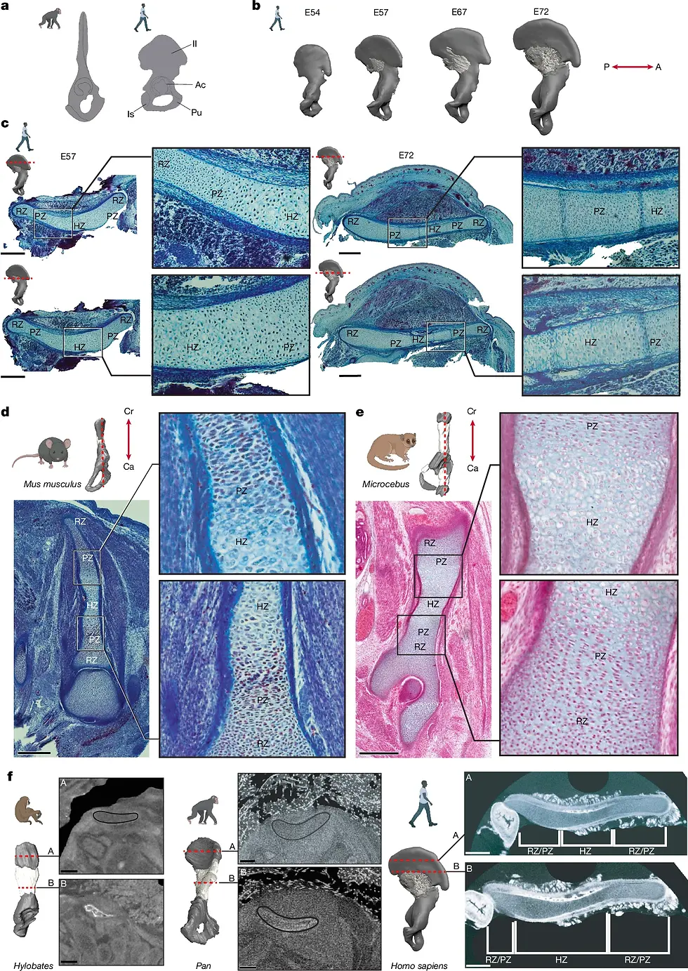
The study examined the ilia of human embryos and early fetuses between approximately gestational days 45 and 72 and compared them with those of laboratory mice (Mus musculus), Microcebus myoxinus, Saguinus geoffroyi, macaques (Macaca), gibbons (Hylobates) and chimpanzees. The findings indicate that the distinctive human ilium arises primarily through two coordinated developmental shifts. First, the direction of cartilage growth changes from longitudinal to transverse. Second, ossification begins at the posterior ilium and advances slowly forwards around its periphery, while the invasion of bone into the interior of the cartilage is substantially delayed.
A growth plate is a region of cartilage that drives skeletal growth during development. Within it, chondrocytes progress through successive stages of rest, proliferation and hypertrophy. As the cells continue to divide, align and enlarge, the cartilage left behind them gradually mineralizes and is replaced by bone. A developing skeletal element therefore extends in the direction in which its growth-plate chondrocytes are organized.


Early in development, the ilium first appears as a cartilaginous model and only later becomes ossified. A mature growth plate contains a resting zone (RZ), proliferative zone (PZ) and hypertrophic zone (HZ). These zones are usually arranged along the long axis of a developing bone, allowing it to lengthen towards both ends. At approximately embryonic day 45 (E45), the human ilium still has a rod-like model aligned along the craniocaudal axis. By E53–E54, however, its chondrocytes begin to proliferate and differentiate rapidly in the anterior–posterior direction. By E72, the growth-plate zones are aligned along the horizontal, or transverse, plane, nearly perpendicular to the original long axis, and expand bidirectionally towards the anterior and posterior sides. This reorientation limits further increase in iliac height while promoting growth in width.
Near the posterior border of the ilium, a small remnant of the original growth plate remains. It is unidirectional, poorly stratified and vertically aligned. Because the iliac growth plates of the non-human primates examined in the study also remained longitudinally oriented, the researchers interpreted this remnant as a developmental trace of the ancestral growth pattern.
Several regulatory genes participate in producing the broad human ilium, including SOX9, PTHLH, which encodes parathyroid hormone-related protein (PTHrP), PTH1R and ZNF521. SOX9 is a transcription factor that directs undifferentiated prechondrogenic mesenchymal cells to become chondrocytes, thereby establishing the initial cartilaginous model of the ilium. At E45, SOX9 is expressed throughout the undifferentiated model. By approximately E53, its expression becomes concentrated at the anterior and posterior ends, whereas the PTH1R receptor is localized mainly to the anterior side, marking the initiation site of the new proliferative zone.
PTHrP signals through PTH1R to maintain chondrocytes in a proliferative state and delay their maturation and hypertrophy. This signalling helps redirect the growth plate from its original craniocaudal orientation into the transverse plane, allowing the ilium to widen anteriorly and posteriorly. The transcription factor ZNF521, expressed in mesenchymal and perichondrial tissues adjacent to the anterior ilium, functions within the same regulatory network as PTH1R and helps control growth-plate size, morphology and expansion. Disruption of these proteins can therefore interfere with transverse iliac growth. Patients carrying mutations in SOX9 or PTH1R, for example, may have markedly narrow ilia with reduced lateral flaring.
In mice and non-human primates, iliac ossification generally begins in the middle of the cartilaginous model. Blood vessels rapidly invade the interior, and bone formation then extends along the longitudinal axis towards the cranial and caudal ends. In humans, iliac ossification begins at approximately E57, but the primary ossification centre forms along the posterior border of each ilium, close to the greater sciatic notch and the sacrum.
New bone initially forms around the outer surface of the cartilaginous model. From its posterior point of origin, the ossification front then advances anteriorly in a distinctive radial pattern. Rather than immediately invading the centre of the cartilage, bone formation remains confined to the peripheral layer for an extended period. This delay allows the ilium to continue growing anteriorly and outwards, ultimately contributing to the broad pelvic form required for bipedal locomotion.

At E72, mineralization and newly formed bone are still largely restricted to the perichondrium, with blood vessels distributed along the external surface rather than penetrating the central hypertrophic cartilage. Internal ossification is delayed by approximately 16 weeks relative to peripheral mineralization and does not begin to extend deeply into the ilium until around gestational week 24, starting near the primary ossification centre.
By retaining cartilage within its interior for such a prolonged period, the ilium preserves tissue capable of continued growth. This permits both anterior expansion and circumferential enlargement while maintaining the complex three-dimensional shape of the iliac blade. The anterior growth zone, which contains the future anterior superior iliac spine and anterior inferior iliac spine, can remain cartilaginous until approximately gestational week 25.
Single-cell data suggest that peripheral osteoblasts may arise from perichondrial cells and subsets of fibroblasts. Once these cells enter the osteoblast lineage, they begin producing bone matrix. This transition may be initiated by RUNX2, whose protein product is a major transcription factor governing the commitment of progenitor cells to an osteogenic programme.
FOXP1 and FOXP2, which become active somewhat later, may regulate the timing and spatial distribution of osteoblast differentiation, allowing ossification to advance slowly and radially along the outer surface of the ilium. At the same time, the angiogenic factors VEGFA and VEGFB are expressed mainly around the iliac periphery, directing blood vessels towards the newly forming peripheral bone. Before E72, the central ilium still lacks strong RUNX2 and VEGF expression. Internal vascular invasion and ossification therefore begin much later than they do in most other developing bones.
These differences in growth and ossification ultimately produce different developmental rates in different parts of the ilium. Posteriorly, the ilium forms a relatively stable connection with the sacrum through the developing sacroiliac joint. Anteriorly, cartilage is retained for longer, allowing the tissue to continue extending and undergoing morphological refinement. This prolonged anterior growth produces structures such as the anterior superior iliac spine and anterior inferior iliac spine.
Human iliac development therefore combines a stable posterior region with an extended period of anterior growth. The resulting ilium is short and broad, curves around the sides of the body and bears several prominent projections that serve as attachment sites for muscles and ligaments.
These projections are particularly important for the transmission of muscular and ligamentous forces. By approximately E53, the gluteus medius and gluteus minimus already attach directly to the posterolateral surface of the cartilaginous ilium. As the iliac blade expands, their areas of origin enlarge laterally. The gluteus maximus is present farther posteriorly at this stage but establishes a clear attachment to the ilium somewhat later.
By approximately E72, the direct head of the rectus femoris has attached to the cartilaginous precursor of the anterior inferior iliac spine. The attachment of the iliofemoral ligament appears considerably later and can be observed by around gestational week 27. After birth, these muscles contribute to hip flexion, extension and abduction. The gluteus medius and gluteus minimus are particularly important during the single-stance phase of walking: when one leg supports the body, they stabilize the pelvis and prevent the unsupported side from dropping.
The researchers further proposed that forces generated by early fetal muscle contractions might feed back into iliac development. Through mechanotransduction, early muscle attachment and contractility could help orient the iliac blade parasagittally and influence the rate at which the anterior perichondral ossification front advances. This possibility remains a hypothesis and has not yet been directly demonstrated.
The researchers then placed these embryological findings within the broader timescale of hominin evolution. Bipedal locomotion involved a suite of anatomical changes, including an inferiorly positioned foramen magnum, an S-shaped lordotic spine, a femoral bicondylar angle and an enlarged, aligned big toe. The fossil record indicates that these traits did not all appear simultaneously. Both Ardipithecus and Australopithecus, for example, already possessed short, broad ilia.
On this basis, the researchers proposed a tentative three-step evolutionary model. During the first stage, approximately 8–5 million years ago, iliac growth shifted—either abruptly or gradually—from a vertical to a transverse orientation through the reorganization of chondrocytes. This change may have accompanied the transition from ape-like locomotion to facultative bipedalism, when greater lateral stabilization of the pelvis became necessary.
During the second stage, approximately 5–2 million years ago, hominins shifted from facultative to obligate bipedalism. Additional molecular changes may have fixed or locked the growth plate into its transverse orientation, leaving only a remnant signal of vertical differentiation. At the same time, the initiation of ossification may have shifted towards the posterior ilium, allowing continued anterior growth in response to changing muscular demands.
During the third stage, beginning around 2 million years ago, the increasing demands of running, together with the evolution of fetuses with larger heads and broader shoulders, may have favoured a further delay in ossification. By extending the period of cartilage growth, this delay would have enabled greater pelvic expansion while preserving the complex shape of the ilium, ultimately contributing to the characteristic form of the modern human pelvis.
Author: Shui-Ye You
References:
Cho JH et al. (2024). Growth plate closure and therapeutic interventions. Clin Exp Pediatr.
Hogervorst T and Vereecke EE. (2014). Evolution of the human hip. Part 1: the osseous framework. Journal of Hip Preservation Surgery.
Senevirathne G et al. (2025). The evolution of hominin bipedalism in two steps. Nature.


Comments