
Large silesaur from Zambia approached 3 metres in length before reaching skeletal maturity

Silesaurs occupy an awkward position in studies of dinosaur origins. They belong to Avemetatarsalia, the bird-line archosaurs, and are very closely related to dinosaurs. Yet their exact evolutionary position remains controversial: they may represent the sister group to Dinosauria, or they may instead form a series of early-diverging ornithischian dinosaurs. Regardless of which hypothesis is correct, the more complete fossils discovered in the past have created a broadly similar impression—silesaurs were generally modest-sized animals. In most known taxa, the femur is no more than 15 cm long, while even the relatively large Silesaurus opolensis had a maximum femoral length of only about 21 cm.
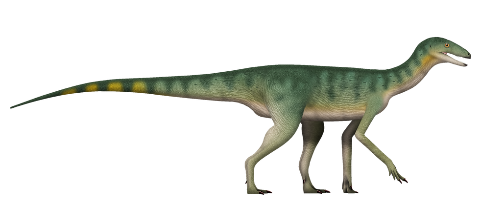
Several fragmentary fossils from East Africa are now changing that picture. One femur, discovered in Zambia in 1963 and subsequently overlooked in a museum collection for decades, indicates that some silesaurs could grow substantially larger. Remarkably, the individual represented by this specimen had not even reached skeletal maturity when it died.
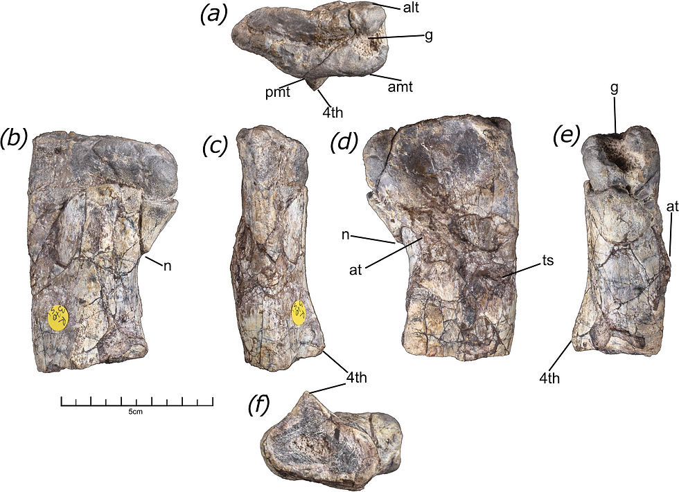
The specimen, NHMUK PV R37051, comes from the Ntawere Formation of Zambia, which is approximately Ladinian to Carnian in age within the Triassic. Only the proximal portion of the left femur, near the hip joint, is preserved, measuring 7.7 cm in length; the distal end is missing. Despite its fragmentary condition, the proximal femur retains several features characteristic of silesaurs, including a deep groove extending across the proximal surface, a notch beneath the femoral head, and a prominent fourth trochanter. These features allowed the specimen to be identified as a silesaur.

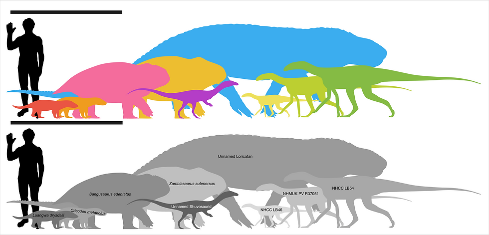
To estimate its original size, the researchers compiled measurements from 59 femora belonging to early-diverging avemetatarsalians and examined the relationship between proximal femoral width and total femoral length. A linear regression was then used to reconstruct the missing length of NHMUK PV R37051. The analysis produced an estimated total femoral length of approximately 26.6 ± 1.8 cm. If the animal had body proportions comparable to those of the more completely known Silesaurus, isometric scaling suggests that NHMUK PV R37051 may have approached 3 m in total body length.
Another femur from the same Ntawere Formation, NHCC LB54, was re-estimated at approximately 33.6 ± 2.7 cm long. Using the same proportional scaling, the animal may have reached around 3.5 m in length. Together with NHMUK PV R16303, a large femur from the Manda beds of Tanzania, there are now three African specimens that substantially exceed the body sizes typical of better-known silesaurs.
The researchers also sectioned NHMUK PV R37051 near the fourth trochanter to examine its bone histology. The cortex contains abundant vascular canals and considerable woven-fibred bone tissue, features associated with relatively rapid bone growth. Closer to the outer surface, vascularization decreases and the tissue begins to transition towards more regularly organized parallel-fibred bone and lamellar bone, indicating that growth was beginning to slow. The bone also shows little secondary remodelling and lacks an external fundamental system, a structure associated with the substantial slowing or cessation of somatic growth.
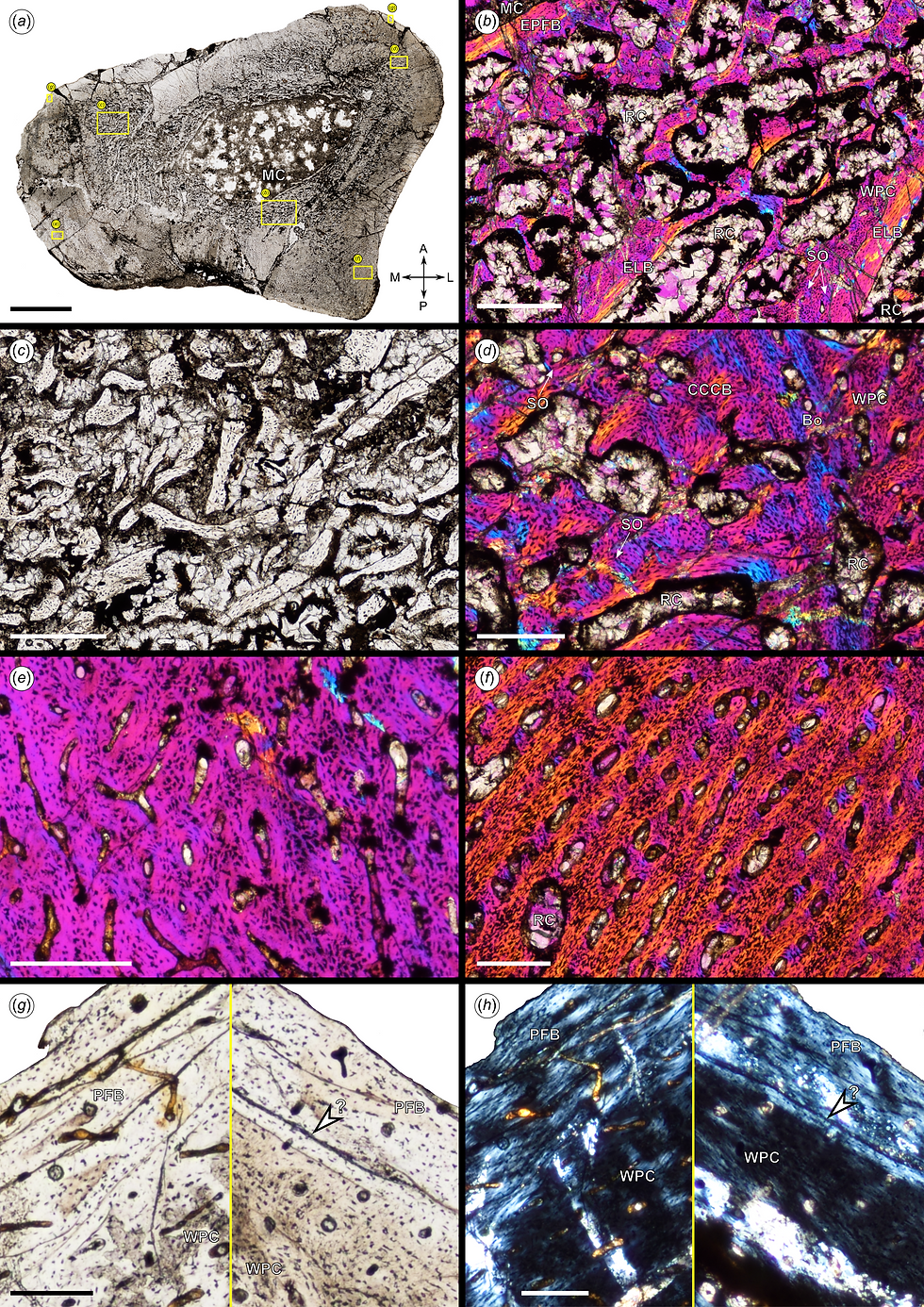
This suggests that the already large animal was still only a late juvenile or early subadult when it died and had not yet finished growing. A possible line of arrested growth is preserved close to the outer surface of the cortex, although cracking and diagenetic alteration make its identification uncertain. If this structure is indeed a genuine growth mark, the individual may have been only around one year old at the time of death.
Two considerably smaller femora, NHCC LB78 and LB79, have also been recovered from the Ntawere Formation. Intriguingly, their bone tissues are dominated by the more slowly deposited parallel-fibred bone, indicating substantially slower growth than in either NHMUK PV R37051 or the large NHCC LB54. If all these femora represented different growth stages of a single species, the smaller individuals would generally be expected to show the faster growth rates associated with earlier ontogeny. Instead, the smaller specimens were growing more slowly while the much larger individuals were still growing rapidly. The striking size differences among Ntawere silesaur femora may therefore reflect the presence of more than one species.
The only formally named silesaur from the Ntawere Formation is Lutungutali sitwensis. Its holotype, however, consists primarily of a pelvic girdle and caudal vertebrae and does not preserve a femur. There is therefore no directly overlapping anatomy that would allow NHMUK PV R37051 to be confidently referred to L. sitwensis. NHMUK PV R37051 also differs in several respects from other silesaur femora recovered from the Ntawere Formation. For example, it possesses a trochanteric shelf, and both the anterior trochanter and fourth trochanter extend farther proximally. These features can vary during ontogeny or among individuals, however, so the differences are not sufficient to justify naming a new species.
Some previous phylogenetic studies incorporated isolated maxillary, femoral and tarsal material from the Ntawere Formation into L. sitwensis, effectively assuming that only a single silesaur taxon was present there. In the new analysis, these unassociated specimens were removed from the character scores for L. sitwensis, restricting the taxon to features that can actually be scored from the holotype. NHMUK PV R37051 was recovered among relatively early-diverging silesaurs and occupied a different position from L. sitwensis. Nevertheless, the femur is extremely incomplete and statistical support for many nodes in the phylogenetic tree is weak, so the analysis cannot yet establish that a second silesaur species was definitely present in the Ntawere Formation.

Silesaurs have often been regarded as relatively minor components of Triassic faunas otherwise dominated by synapsids and pseudosuchians. In the Ntawere Formation, however, silesaur remains are known from at least seven or eight localities, including one bonebed containing at least seven individuals. Their fossils therefore indicate that silesaurs were neither rare members of this ecosystem nor exclusively small-bodied animals.
Author: Shui-Ye You
Reference:
Lovegrove J et al. (2025). A new large 'silesaur' specimen from the ?Late Triassic of Zambia; taxonomic, ecological and evolutionary implications. R. Soc. Open Sci.


Comments