
The Painted "Skin" of a Permian Reptile—The Forged Fossil of Tridentinosaurus antiquus

In 1931, an unusual reptile fossil was discovered in the Italian Alps. At the centre of the slab was the clearly visible outline of a slender, sprawling animal, its body extending from the trunk and limbs into a long tail. The dark-brown body outline contrasted sharply with the surrounding pale-pink tuffaceous sandstone. The specimen was later named Tridentinosaurus antiquus.

When the specimen was formally described in 1959, the dark-coloured outline was interpreted as skin and other soft tissues preserved through carbonization. If genuine, such preservation would have been exceptionally rare. Early Permian deposits in the Alps have yielded numerous tetrapod trackways, but body fossils are considerably scarcer, and skeletons preserving evidence of soft tissues are rarer still. Yet although Tridentinosaurus appeared remarkably complete at first glance, very few of its bones could actually be identified.
In the hindlimbs, the femora, tibiae, and fibulae are barely recognizable, while most of the remaining skeleton is poorly defined and the skull is completely absent. Earlier interpretations of the animal's body proportions, neck length, limb shape, and trunk outline therefore relied heavily on the dark-coloured material surrounding the bones. Tridentinosaurus was assigned to different groups of early reptiles over the years and was later placed within the polyphyletic group Protorosauria. For decades, the material that appeared to represent its "skin" played an important role in determining the animal's identity.
In 2024, Valentina Rossi and colleagues re-examined the specimen and noticed a feature inconsistent with the expected preservation of an organically preserved compression fossil: the animal's outline was not flat. Vertebrate soft tissues preserved as compression fossils are typically flattened during burial and generally show little surface topography. A reconstructed 3D surface model of Tridentinosaurus, however, revealed that the hindlimbs and tail stand noticeably higher than the abdomen and forelimbs, while a raised ridge runs through the centre of the abdominal region. Mechanical preparation marks are also visible around the specimen, particularly near the forelimbs and abdomen. The researchers interpreted much of this uneven topography as the result of extensive preparation, probably carried out in an attempt to expose more of the skeleton.

Another clue emerged when the fossil was examined under ultraviolet light. The dark body outline, together with parts of the bones, fluoresced yellow. Organically preserved soft tissues in vertebrate fossils do not normally show this response, whereas lacquers, varnishes, glues, coatings, and some artificial pigments can fluoresce under ultraviolet light. A fossilized plant fragment recovered from the same geographical area and preserved in similar rock was subjected to the same test, but did not produce comparable fluorescence.
The microscopic structure of the dark material raised further doubts. The researchers removed tiny samples from several parts of the body outline and examined them using scanning electron microscopy. Instead of structures typical of preserved soft tissues, they found a layer composed of numerous angular granules embedded within a microcrystalline matrix. These particles ranged from less than 2 μm to about 20 μm across. Elemental analysis showed that many of the granules were enriched in calcium and phosphorus, while X-ray diffraction revealed abundant apatite together with quartz, feldspar, and phyllosilicates also present in the surrounding rock matrix.

None of these observations alone was enough to demonstrate that the specimen had been forged. More than 200 million years of burial and diagenesis can profoundly alter genuine biological tissues. The researchers therefore turned to Raman spectroscopy and attenuated total reflectance Fourier-transform infrared spectroscopy to examine the chemistry of the dark material more directly. Raman spectroscopy detected two broad bands at approximately 1350 and 1580 cm⁻¹, corresponding to the D and G bands commonly associated with disordered carbon. Such signals can occur in melanin, but they can also be produced by coal, kerogen, charcoal, and manufactured carbon-based pigments. The Raman results by themselves therefore could not distinguish fossilized biological material from artificial pigment.
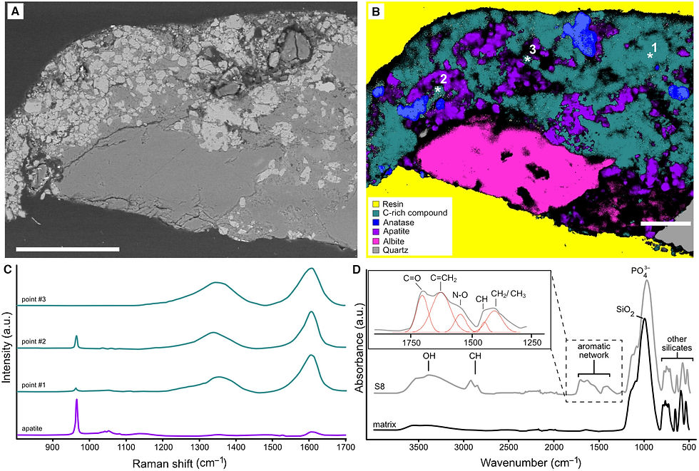
To resolve this problem, the team compared the material from Tridentinosaurus with melanosomes extracted from the skin of a living reptile and with commercially available bone black pigment. The spectrum obtained from the dark material in the abdominal region did not show the chemical signature expected for fossilized melanin. Instead, it more closely resembled a manufactured carbon-based black pigment. The dark layer also contained chemical signals compatible with organic binding materials.
Taken together—the ultraviolet fluorescence, granular microscopic texture, abundance of apatite, lack of recognizable melanosomes, and spectroscopic evidence—the results strongly indicated that the body outline was made from a manufactured carbon-based pigment applied to the surface of the rock. The researchers identified it as most consistent with bone black paint mixed with an organic binder. One or more additional coatings may also have been applied over the specimen.
Some genuine fossil bones were apparently present in the rock from the beginning. At some point, the surrounding slab was mechanically prepared and dark pigment was applied around these remains, connecting the fragmentary bones into the outline of a slender, lizard-like animal.
Who altered the fossil remains unknown, and there is no evidence establishing why it was done. The museum has no complete record of the specimen's early preparation or conservation history. What can be inferred is that the alteration predates the formal 1959 description, because by then the dark body outline was already being treated as an original part of the fossil.
The discovery creates another problem: is Tridentinosaurus antiquus itself still a valid taxon?
Many of the characters originally used to establish and classify the animal—including features of the neck, abdomen, forelimbs, hands and feet, and overall body proportions—were derived partly or entirely from the artificial outline. The genuine long bones of the hindlimbs appear to be authentic fossils, but they are poorly preserved and lack diagnostic features such as processes and foramina that would normally be useful for taxonomic comparison. The forelimbs and girdles cannot be reliably identified, no clear vertebrae are visible in the neck or tail, and the skull is absent. Even along the fractured surface where the head should have been located, no bone cross-sections attributable to the skull could be identified.
At present, the main anatomical information that can still be used for comparison is limited to the approximate proportions of the femora, tibiae, and fibulae. The researchers did not formally declare Tridentinosaurus antiquus invalid, but they advised caution in using the specimen in phylogenetic analyses of early reptiles. Modern tomographic techniques may eventually reveal whether additional skeletal remains are still hidden inside the rock. Until then, the true identity of this enigmatic Permian animal remains unresolved.
Author: Shui-Ye You
Reference:
Rossi V et al. (2024). Forged soft tissues revealed in the oldest fossil reptile from the early Permian of the Alps. Palaeontology.


Comments